
CMAC National Facility - X-Ray Diffraction and Solid-State Characterisation
Definitive structural insight to de-risk development
Unanswered solid-state questions pose risks for pharmaceutical developers and materials researchers. They can slow down decisions, complicate formulations and weaken regulatory confidence.
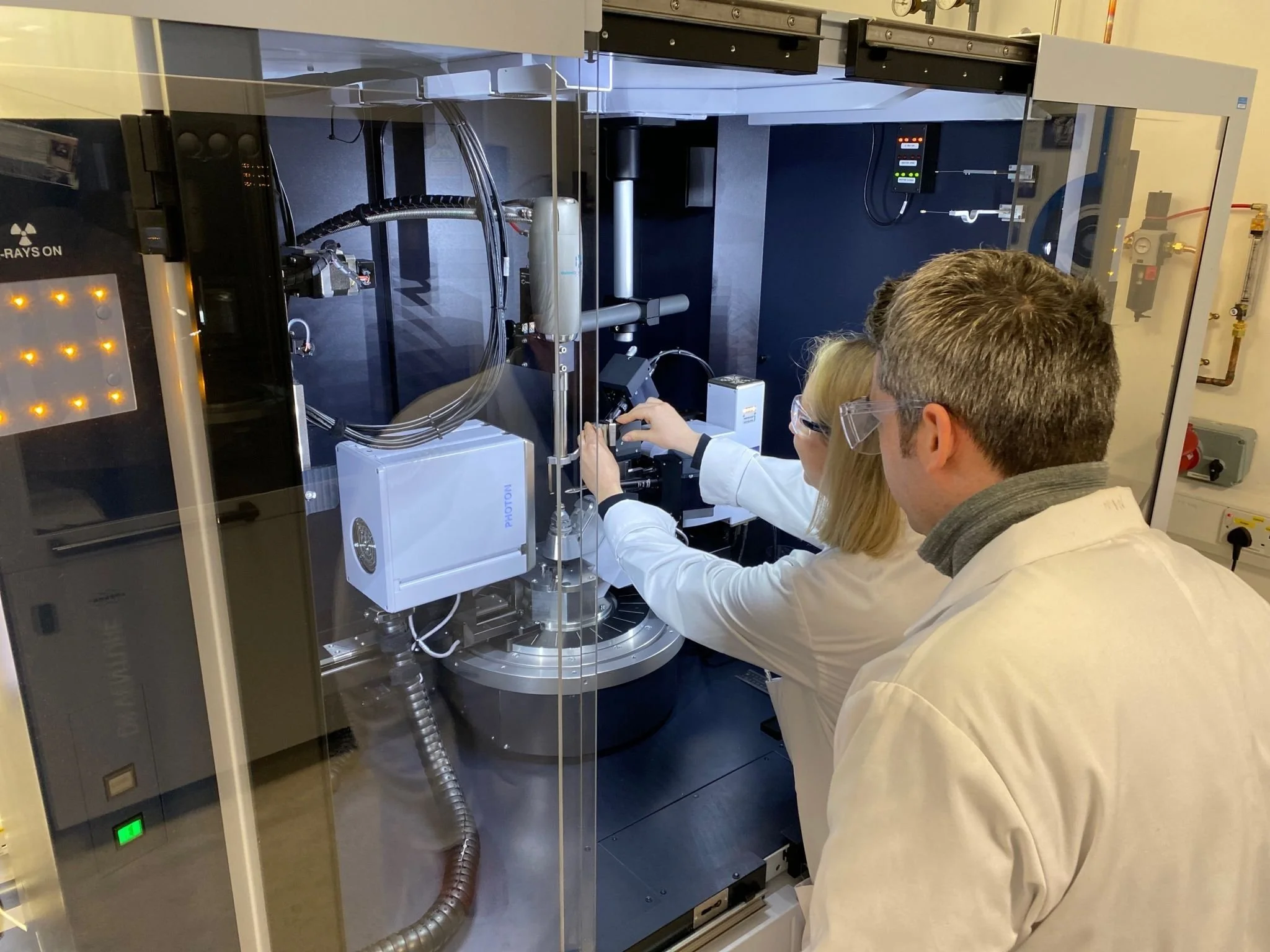
WHAT SETS US APART
A complete solid-state landscape in one place
Our integrated analytical and experimental environment maps the entire solid-state landscape — from single crystals to amorphous materials.
Expert-led, problem-focused studies
Our crystallographers and solid-state specialists connect structure, stability, transitions and microstructure directly to product performance.
ANALYTICAL PLATFORMS
Seven techniques. One integrated facility.
-
Identify what’s in your sample, and how much. Powder X-ray diffraction is the gold standard for determining crystalline phases in complex materials — matching diffraction patterns against extensive reference databases to rapidly identify phases present and quantify their relative proportions, supporting manufacturing troubleshooting, crystallisation monitoring and product consistency verification.
Where single crystals aren’t available, we go further: structure determination directly from powder data is a highly specialised capability available at only a small number of facilities worldwide. This lets you resolve ambiguous solid forms with confidence, support patent, IP and regulatory arguments, and advance development without dependency on crystal growth.
Capabilities: phase identification and quantitative analysis; reflection and transmission (capillary, Cu Kα1) geometries; high-throughput multi-well plate screening; ab initio structure solution from powder data.
-
Understand how your materials respond to real-world conditions. Using controlled temperature, humidity, gas flow or vacuum, we monitor structural transformations including polymorphic transitions, hydrate formation, desolvation and thermal phase changes. Providing critical insight into material stability under realistic storage and processing conditions.
We also carry out high-pressure powder diffraction in-house. A niche capability, it reveals polymorphs and phase transitions under compression that stay hidden at ambient pressure. Paired with our in-house high-pressure single-crystal work, this gives you compression studies across both powders and single crystals, not widely available elsewhere.
Capabilities: variable temperature and humidity diffraction; in situ high-pressure powder diffraction; formulation and manufacturing strategy support.
-
Get complete crystal structures with full 3D details for small molecules, salts, crystals, solvates and complex multi-component systems. This allows for confident polymorph identification, analysis of hydrogen-bonding networks, and interpretation of physicochemical properties for pharmaceutical development and regulatory documentation.
Our in situ high-pressure crystallography uses diamond anvil cells to study structures under compression. This capability is not widely available elsewhere, giving you access to polymorphs, phase transitions and pressure-driven structural changes that stay hidden at ambient conditions.
Capabilities: Cu and Mo X-ray sources; precise temperature control (80–400 K); in situ high-pressure crystallography via diamond anvil cell (ambient–10 GPa).
-
Explore local and short-range order in materials that conventional diffraction can’t reach. xPDF analysis reveals structural insight in poorly crystalline phases, nanomaterials and disordered or amorphous pharmaceutical materials. This includes temperature-dependent studies of relaxation and amorphous–crystalline transitions.
Capabilities: Mo-source total scattering; wide Q-range; variable temperature control (80–500 K).
-
Visualise internal structures and microstructure without sectioning or sample preparation. Micro-computed tomography (microCT) provides non-destructive three-dimensional imaging at micron-scale resolution. This enables quantitative characterisation of porosity, inclusions, defects and particle morphology through advanced 3D image analysis.
Capabilities: Bruker SkyScan 2214, ~300–500 nm resolution; in situ heating, cooling and mechanical testing.
-
Explore structural organisation from the nanoscale up. Simultaneous SAXS and WAXS measurements link nanoscale organisation with crystalline structure, supporting particle size, shape and aggregation analysis across formulations and devices under in situ temperature, humidity or shear conditions.
Capabilities: Xenocs Xeuss 2.0; 0.1–100 nm range; nanoparticle and polymer characterisation.
-
Get structure when neither single crystals nor useful powder data are available. Electron diffraction extends our X-Ray capabilities to microcrystalline and nanocrystalline materials. It helps us to characterise pharmaceutical solid forms like polymorphs, salts, cocrystals and solvates especially when single-crystal X-Ray diffraction is not possible.
Individual crystallites can be examined directly from heterogeneous or multicomponent samples, including material isolated from powders and ballmilled mixtures, with automated crystal detection supporting screening across multiple crystallites in a single sample.
Capabilities: cryogenic data collection for sensitive, hydrated or solvated materials; automated crystal detection and multi-dataset comparison; three-dimensional structure solution and refinement from very small crystals.
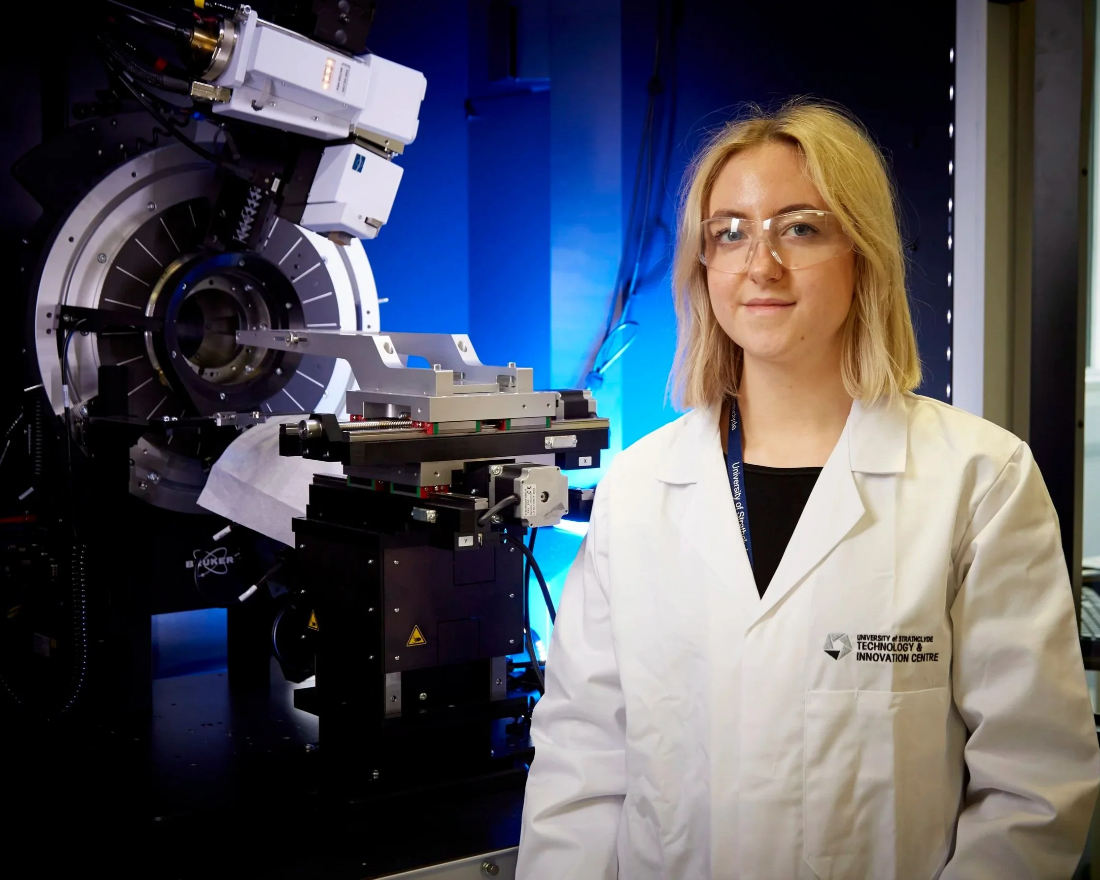
Not sure which platform you need?
Our experts will advise on the best approach for your question and material.
APPLICATIONS AND IMPACT
Our analytical suite supports
Solid-form selection, polymorph control and stability studies
Structure–performance relationships
Nanostructure and microstructure evaluation
Drug product development
Amorphous and poorly crystalline materials
Materials research in pharmaceuticals and beyond
Ready to collaborate?

Whether you're exploring new drug products or solving complex process challenges, we are here to help.

CMAC has achieved two Silver and one Bronze level certifications for integrating sustainable practices in its lab operations.