
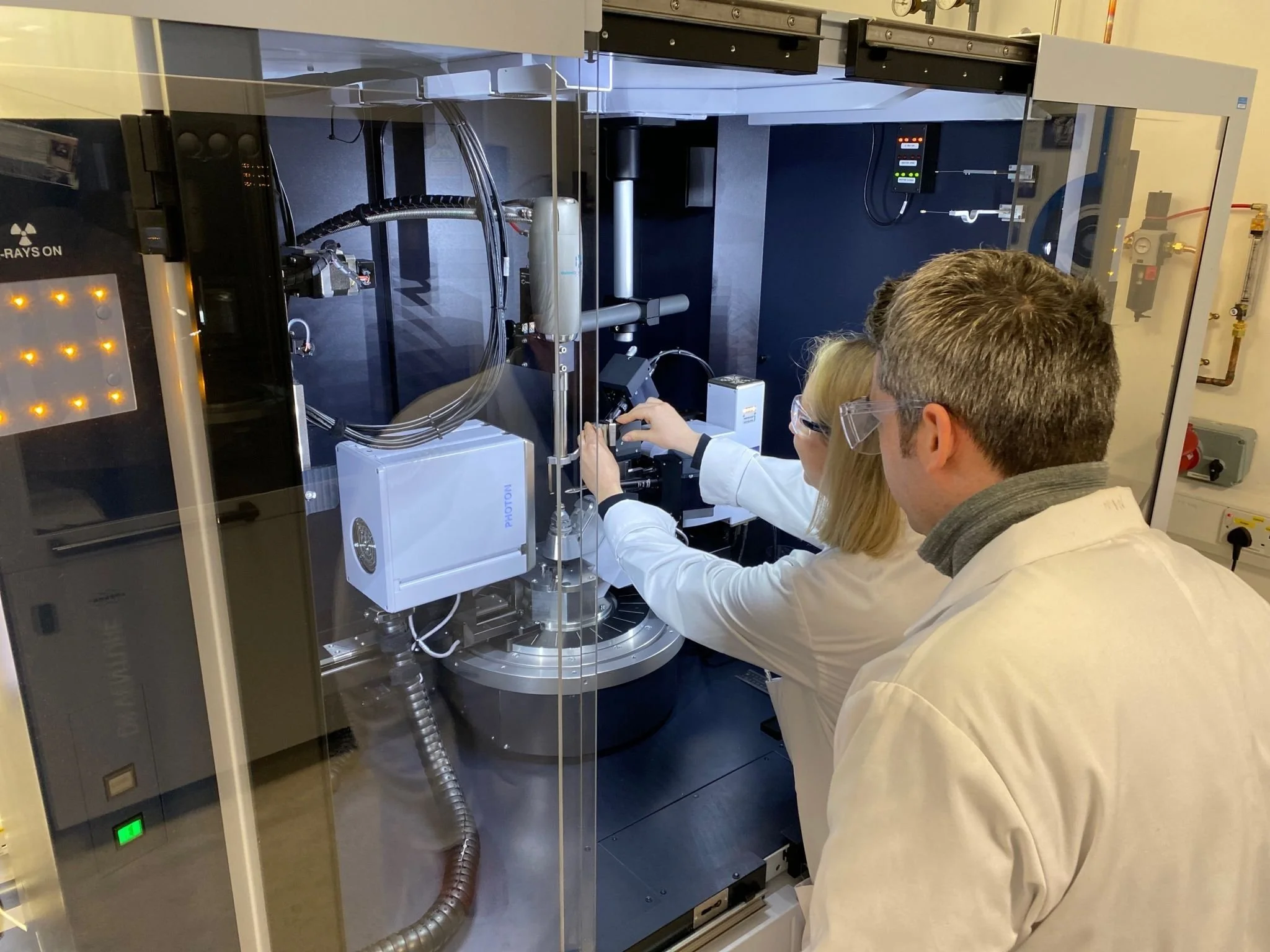
CMAC National Facility - X-Ray Diffraction and Solid-State Characterisation
Definitive structural insight to de-risk development
For pharmaceutical developers and materials researchers, unanswered solid-state questions create risk - slowing decisions, complicating formulations and undermining regulatory confidence.
Our integrated hub enables you to de-risk solid form selection early in development to:
Make faster, evidence-based decisions on polymorphs, salts and form selections
Strengthen regulatory submissions with structural justification
Characterise and solve challenging solid forms where single crystals are unavailable
Avoid late-stage reformulation and stability surprises.
We focus on outcomes - not just measurements to ensure data translates into decisions.
What sets us apart

Structure solution from powder data - a rare capability
Structure determination from powder X‑Ray diffraction data is a highly specialised capability, available at only a small number of facilities worldwide.
At CMAC, we routinely solve structures directly from powder data, supporting development programmes where single‑crystal material cannot be obtained or where timelines don’t allow repeated crystallisation attempts.
This allows you to:
Resolve ambiguous solid forms with confidence
Support patent, IP and regulatory arguments
Advance development without dependency on crystal growth
Reduce uncertainty in solid‑state risk assessments
This capability is particularly valuable in early‑stage pharmaceutical development, where material is limited and decisions are time‑critical.
-
A complete solid-state landscape in one place
Our integrated analytical and experimental environment allows us to map the entire solid‑state landscape, from single crystals to amorphous materials.
-
Expert-led, problem‑focused studies
Our crystallographers and solid‑state specialists deliver studies that connect structure, stability, transitions and microstructure to product performance.
Core capabilities:
-
Identify what’s in your sample, and how much.
Powder X-Ray diffraction is the gold standard for determining crystalline phases in complex materials. By matching diffraction patterns against extensive reference databases, we rapidly identify the phases present and quantify their relative proportions.
These capabilities support troubleshooting manufacturing issues, monitoring crystallisation outcomes, verifying product consistency and analysing unknown materials.
-
Understand how your materials respond to real-world conditions.
Using controlled temperature and humidity diffraction experiments, we monitor structural transformations including polymorphic transitions, hydrate formation, desolvation and thermal phase changes.
These measurements provide critical insight into material stability under realistic storage and processing conditions, supporting form stability assessment, identifying transformation pathways and informing formulation and manufacturing strategies.
-
Get complete crystal structures - even without crystals.
We provide full three-dimensional structure determination using state-of-the-art single-crystal and powder X-Ray diffraction. When suitable single crystals cannot be obtained, advanced computational methods allow us to solve structures directly from high-quality powder data.
This capability spans small molecules, salts, cocrystals, solvates, hydrates and complex multi-component systems. Structural insight enables confident polymorph identification, understanding of hydrogen-bonding networks and molecular packing, and interpretation of physicochemical properties, supporting pharmaceutical development, materials research and regulatory documentation.
-
Visualise internal structure without sample preparation.
X-Ray microscopy and computed tomography provide non-destructive 3D imaging at micron-scale resolution, revealing pores, inclusions, cracks and phase distributions all without altering your sample.
This technique offers insight into particle morphology, internal architecture and structural heterogeneity across pharmaceutical, advanced materials and device applications. Combined with diffraction-based methods, it links 3D microstructure to crystallographic and compositional data to give you a full picture of your material from the inside out.
-
Explore structural behaviour under extreme conditions.
High-pressure X-Ray diffraction reveals how materials respond to compression, uncovering new polymorphs, phase transitions and pressure-driven structural changes that remain hidden at ambient conditions.
Using diamond anvil cells and other specialised equipment, we access regions of the solid-form landscape that offer unique insight into intermolecular interactions, packing efficiency and structural stability. This capability is particularly valuable for fundamental research and advanced solid-state investigations.
High‑Performance X‑Ray & Solid-State Platforms
-
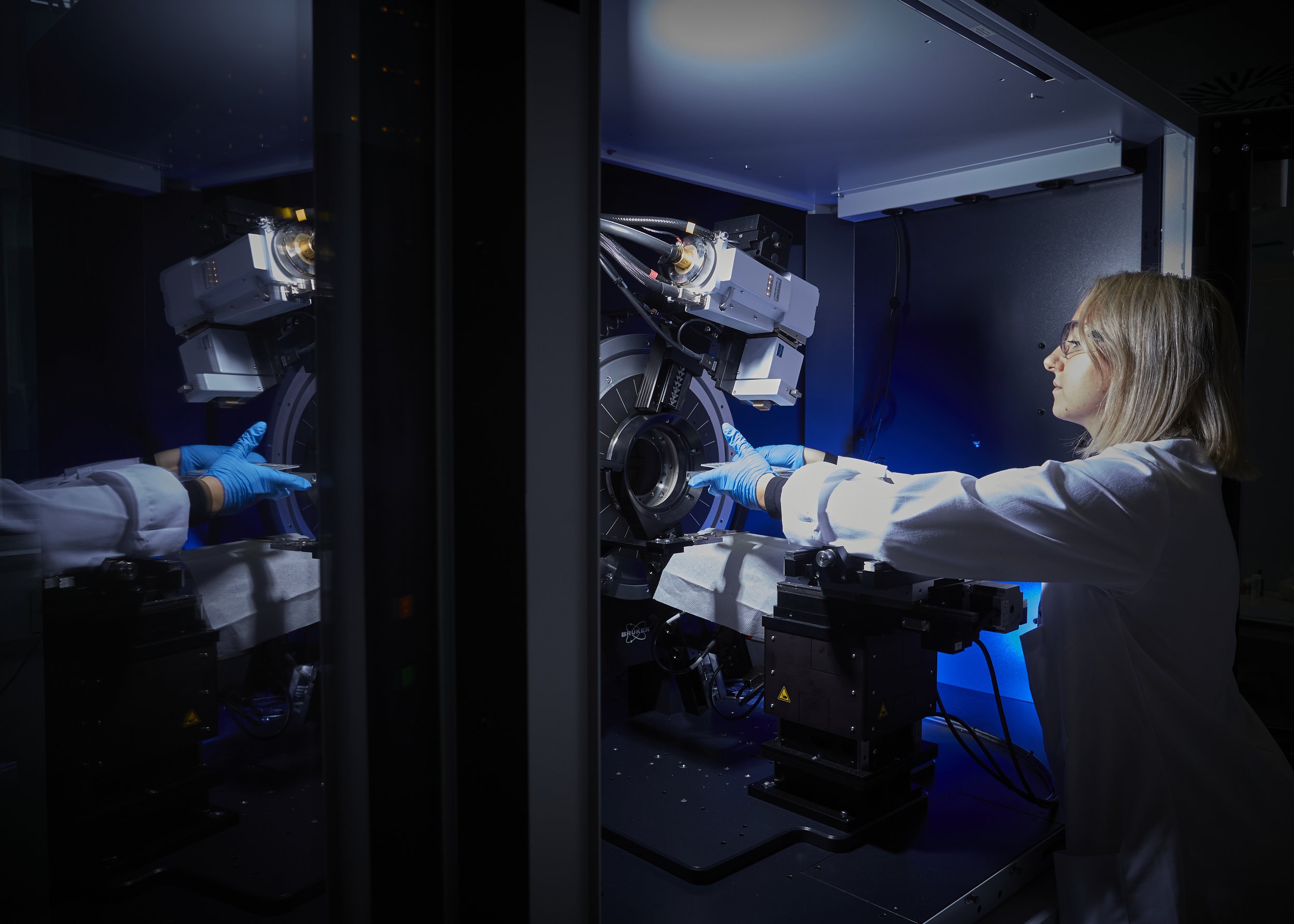
Single crystal X-Ray diffraction
High-quality single crystal structure determination for small molecules, salts, cocrystals, solvates & polymorphs.
Temperature-dependent studies, & custom crystallographic services, including data collection, refinement, and collaborative projects.
Cu and Mo X-ray sources with precise temperature control (80–400 K), supporting challenging samples and weakly diffracting crystals.
Insitu High pressure crystallography (Diamond anvil cell; pressures ca. ambient – 10 GPa).
-

Powder X-Ray diffraction
Phase identification, phase purity assessment, quantitative phase analysis, and ab initio structure solution from powder data.
In situ and non-ambient PXRD under controlled temperature, humidity, gas flow, or vacuum to study phase transformations and stability.
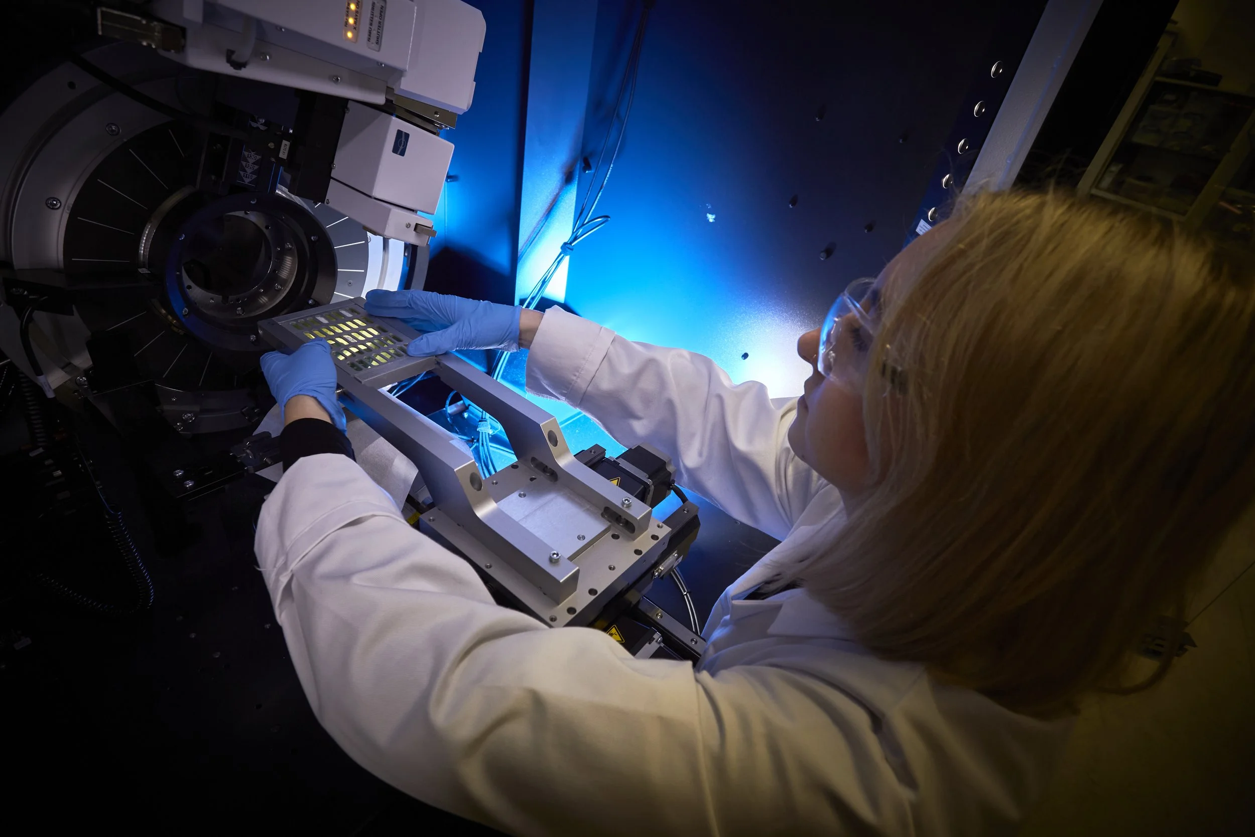
High-throughput screening using multi-well plates for rapid data collection.
Reflection and transmission (capillary, Cu Kα₁) geometries enabling surface-sensitive measurements and high-resolution structural analysis of pharmaceutical solids.
-
X-Ray pair distribution function (xPDF)
Mo-source provides total scattering measurements, wide Q-range, & variable temperature control (80–500 K).
Pair distribution function (xPDF) analysis revealing local and short-range order in crystalline, nanocrystalline, and amorphous pharmaceutical materials.
Powerful for studying poorly crystalline phases, nanomaterials, disorder, and early-stage solid forms inaccessible by conventional PXRD.
Temperature-dependent xPDF studies to probe structural changes, relaxation, and amorphous–crystalline transitions.
-
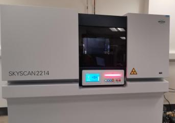
X-Ray nano-computed tomography (nano-CT)
Bruker SkyScan 2214 provides non-destructive 3D imaging of materials, from sub-micron features to whole devices.
High-resolution scanning (~300–500 nm), multiscale analysis, and quantitative assessment of morphology, porosity, particle size, cracks, and defects.
In situ options include heating, cooling, and mechanical testing.
Ideal for studying pharmaceuticals, composites, medical devices, low-density or multi-material systems, and failure analysis, giving detailed insights across a range of sample sizes.
-
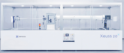
Small and wide-angle X-Ray scattering (SAXS/WAXS)
Xenocs Xeuss 2.0 SAXS/WAXS.
Quantitative analysis of particle size, shape, aggregation, and internal organisation across length scales from ~0.1–100 nm.
Simultaneous SAXS and WAXS measurements for hierarchical structural analysis, linking nanoscale organisation with crystalline structure.
In situ environments (temperature, humidity, shear/flow) supporting dynamic studies of formulations and devices.
Nanoparticle and polymer characterisation, domain spacing analysis, extended q-range measurements and XRD mapping for nanostructure and crystalline domains.
Applications and impact.
Our analytical suite supports:
Solid-form selection, polymorph control and stability studies
Nanostructure and microstructure evaluation
Amorphous and poorly crystalline materials
Drug product development
Structure performance relationships
Materials research in pharmaceuticals and beyond
Get in touch